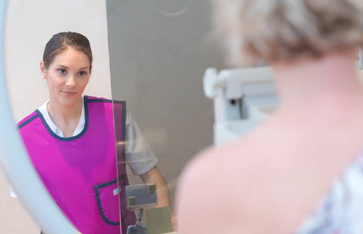
Is a 3D Mammogram a Good Idea?
Research shows there are advantages, especially for older people
As part of breast cancer prevention, the American Cancer Society recommends getting a mammogram annually between the ages of 45 and 54, then every two years from age 55 onward -- although, most doctors involved in breast cancer recommend a yearly mammogram after the age of 40.

Today, many health care organizations offer both 2D and 3D mammography, though it's very possible a patient might not know what type of mammogram they're getting.
Two-dimensional mammography has been available for decades, but in 2011, the Food and Drug Administration (FDA) approved 3D mammography (also known as digital breast tomosynthesis). Since then, a growing number of facilities nationwide have offered 3D mammography to patients, sometimes combining 2D and 3D into one exam.
Because the technology is relatively new, and because 3D mammography isn’t available everywhere yet, a patient may not know enough about it to request one or the other.
Beneficial for Older People
A recent study by researchers at Massachusetts General Hospital shows that the newer exam is beneficial for older people: They found that 2D and 3D mammography detected cancers at the same rate, but 3D mammography detected fewer lymph-node-positive breast cancers in women 65 and older, suggesting that this method discovers cancer at an earlier stage.
"A 3D mammogram detects more invasive cancers and leads to fewer false positive exams than 2D mammography."
The study also showed that 3D mammography resulted in fewer false positives in women 65 and older. For these reasons, 3D mammography may appeal to older people.
“3D mammography is considered to be the ‘better mammogram,’” says the study’s author Dr. Manisha Bahl, director of the breast imaging fellowship program at Massachusetts General Hospital.
“It detects more invasive cancers and leads to fewer false positive exams than 2D mammography,” she continues. “I believe that these benefits of 3D mammography outweigh the risk of increased radiation dose, as the radiation dose of combined 3D and 2D mammography is relatively low and remains below the acceptable limit.”
Differences in Radiation Exposure
Research by scientists at the University of Pennsylvania found that the images acquired from 3D mammography resulted in a 25.6% increase in mean average glandular radiation dose (the average dose absorbed to the breast glandular tissue) than during 2D mammography. (The researchers only looked at one 3D system model; several are available.)
People may be concerned about increased radiation exposure from 3D mammography, but this is unwarranted, according to the researchers.
“Given the benefits that 3D mammography provides compared to 2D mammography, radiation dose exposure should not be a major concern,” says Bruno Barufaldi, a postdoctoral researcher at Penn Medicine in the University of Pennsylvania Health System. “We do not acquire X-ray breast images for cosmetic purposes; there is a medical need behind screening with 3D mammography, which has been saving thousands of patient lives per year.”
Before getting a 3D mammogram, a patient can inquire about the exam. There are two methods: One takes two sets of images, 2D and 3D, which would expose a patient to more radiation.
The other uses 3D mammography alone, then has a computer use the 3D data to create 2D images. This is done because some physicians and health care organizations believe it’s important to look at both 3D and 2D images for a comprehensive exam.
“That is becoming much more common, and then it is really no more dose than your original 2D mammogram,” says Dr. Cheryl Williams, associate professor of radiology and lead interpreting physician for mammography at the University of Nebraska Medical Center. “But even if it’s a double dose, it’s well within the standards set by the FDA, and it’s still an incredibly minimal amount of X-ray beams. It’s nothing like getting a CT (computerized tomography) scan done.”
Similar-Looking Machines and Methods
A patient may not be able to tell from an exam alone which type of mammogram was done.
“For the patient, the exam is nearly identical, because the breast is compressed in a similar fashion,” says Dr. Stefanie Woodard, a breast imager at the University of Alabama at Birmingham. “With 3D, the machine rotates in an arc around the breast and takes multiple pictures as it turns. This allows for detection of more subtle lesions which may only be identified in one of these angles.”
The resulting images from 2D versus 3D mammograms are markedly different, with 3D images showing many “slices” of the breast from different angles. If you were looking for one black seed in a seedless watermelon, you’d spot it more easily if you cut the melon into slices, and that’s what 3D images do.
“This is more like a CT scan, where you slice through the body,” Williams says.
Costs for 3D mammography vary by the facility, but it may only be $50 to $100 more than 2D mammography. Medicare covers 3D mammography, and health insurance companies have different policies about payment.
“Insurance coverage is also variable but seems to be changing in support of 3D mammography,” Woodard says. “This makes sense from a financial standpoint, as these patients will, overall, have less imaging and less costs incurred due to better characterization of the breast tissue.”
The machines are more expensive, but a growing number of facilities, nationwide, are offering 3D mammography.

